Binding Sites and Reactivity
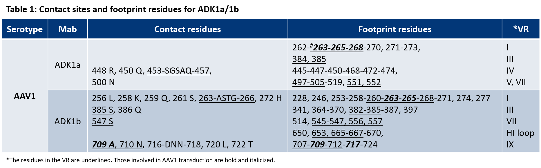
The ADK1a antibody used for the AAV1 Titration ELISA as well as the ADK1b specifically recognize assembled AAV1 capsids, hence epitopes that result from a specific conformational assembly of the capsid proteins VP1, VP2 and VP3. In the publication cited below the contact sites and footprint residues identified for ADK1a and ADK1b are described and the results are summarized in table 1. Multiple contact sites and footprint residues have been identified, that are very likely to be part of the binding site. The amino acids of each binding site are located in different parts of the protein chains and are recognized as the epitope of the antibody only in the assembled capsid where they are in close proximity to each other and in the correct conformation.
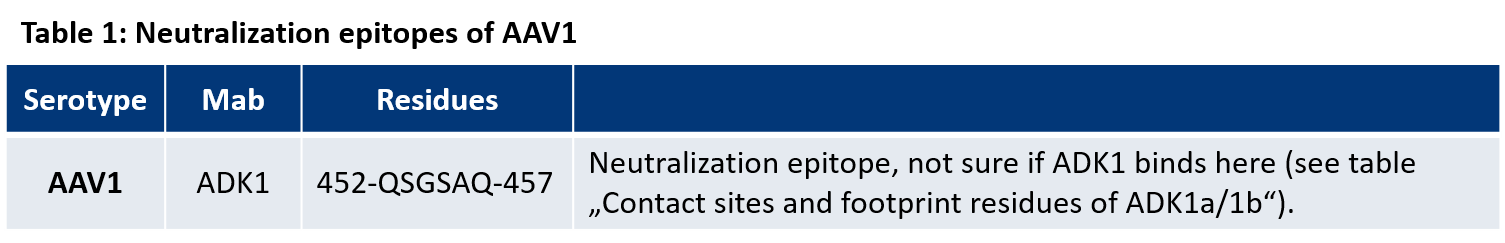
For AAV1 the neutralization epitope has been identified and might give an additional indication about the binding residues of the neutralizing antibody ADK1 used for the corresponding AAV1 Titration ELISAs.
Find more information on the neutralizing activity of the ADK1 antibody in the following publication:
Tseng, Y.-S. et al. Adeno-Associated Virus Serotype 1 (AAV1)-and AAV5-Antibody Complex Structures Reveal Evolutionary Commonalities in Parvovirus Antigenic Reactivity. J. Virol. 89, 1794–1808 (2015).
 mouse monoclonal, ADK1a, lyophilized, purified, sample")
- Purified, lyophilized
- Mouse monoclonal
- Suitable for dot blot, ELISA, ICC/IF and neutralization assay
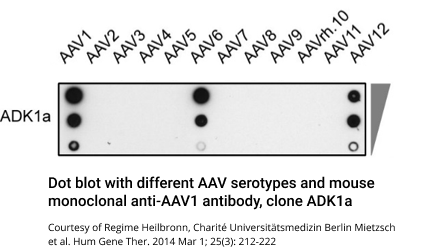
- Reacts with AAV1, AAV6, AAV12 intact capsids
- Isotype: IgG2a lambda
 as detection antibody (1:80)")
- Biotin conjugate
- Mouse monoclonal
- Suitable for dot blot, ELISA, ICC/IF and neutralization assay
- Reacts with AAV1, AAV6, AAV12 intact capsids
- Isotype: IgG2a lambda