AAV8 VP3, recombinant protein
Key Features
• Recombinant AAV8 capsid protein VP3
• Useful in combination with AAV8 VP1 and VP2 for ratio analysis (also available as set, Cat. No. 72008)
• Precise molar concentration
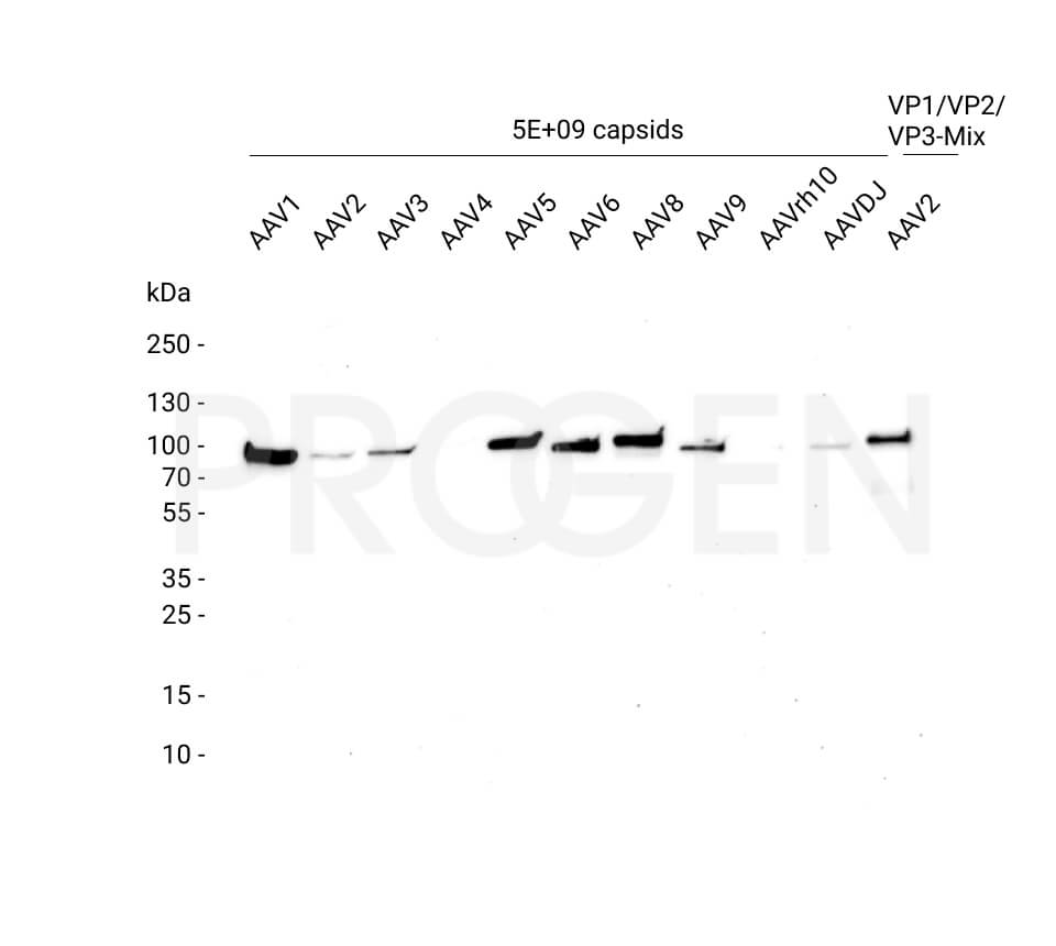
• Suitable for WB, dot blot, SDS PAGE
• Useful in combination with AAV8 VP1 and VP2 for ratio analysis (also available as set, Cat. No. 72008)
• Precise molar concentration
• Suitable for WB, dot blot, SDS PAGE
Not available in China and in the Russian Federation.
For US shipment, the packages are only sent out for delivery on Mondays and Tuesdays, in the EU from Monday until Wednesday.
Product description
| Quantity | 10 µg |
|---|---|
| Application | Dot blot, SDS PAGE, WB |
| Purification | Ni-NTA chromatography |
| Storage | -80°C |
| Intended use | Research use only |
| Concentration | 100 µg/ml (1.61 µM) |
| Formulation | Liquid, 6 M urea in PBS |
| Source | Escherichia coli |
| Molecular weight | 62.1 kDa (calculated Mw from aa sequence) |
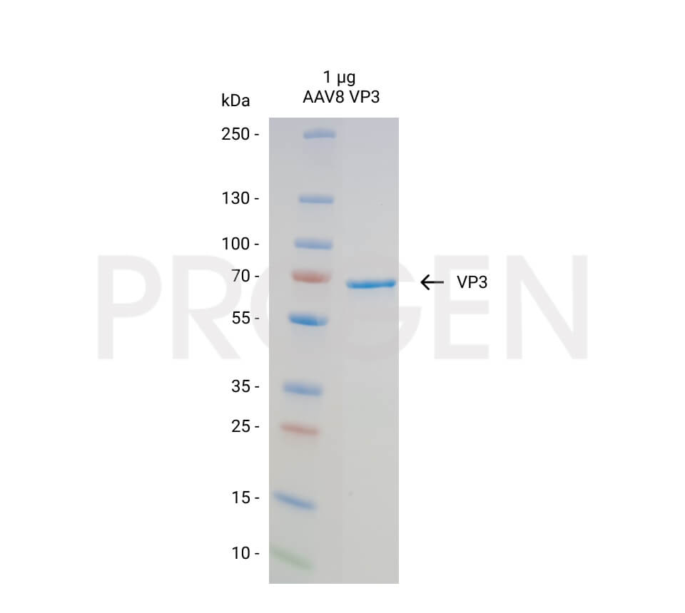
| Purity | > 95% (determined by SDS PAGE) |
| Product description | N-terminal His-tagged (MGSSHHHHHHSSGLVPRGSH) recombinant AAV8 capsid protein VP3 |
Applications
| Tested applications | Tested dilutions |
|---|---|
| Dot Blot | 100 ng, depending on primary antibody and detection method |
| SDS PAGE | 1 µg |
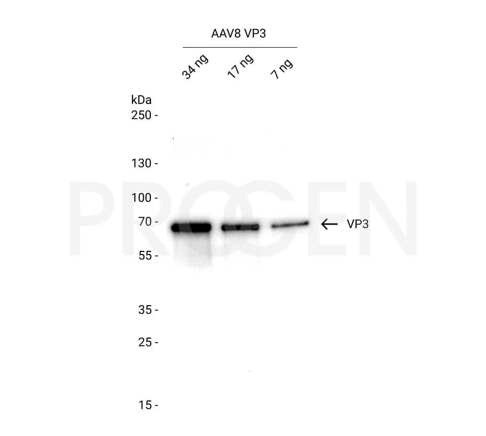
| Western Blot (WB) | 5-20 ng, depending on primary antibody and detection method |
Background
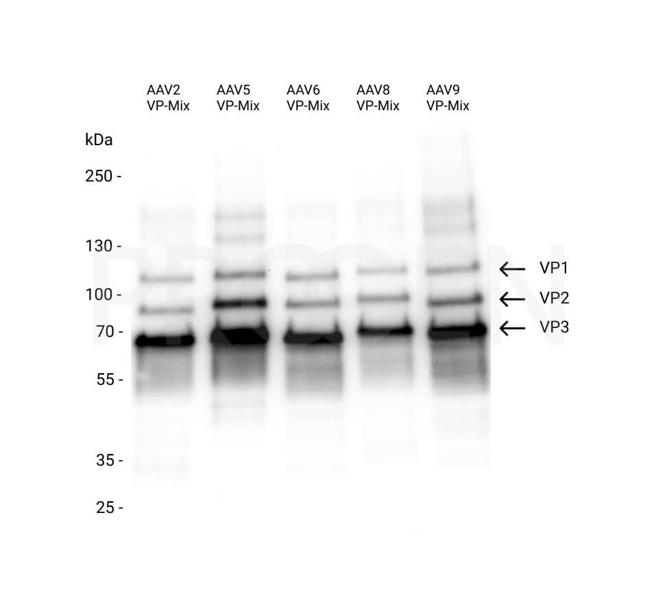
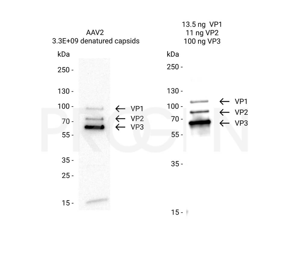
The AAV capsid consists of three capsid proteins, i.e. VP1, VP2 and VP3, which differ in their N-terminus and encapsulate the genomic ssDNA. In native virus particles, the three proteins form subunits with a ratio of 1:1:10 (VP1:VP2:VP3), in a total number of 60 subunits per capsid.
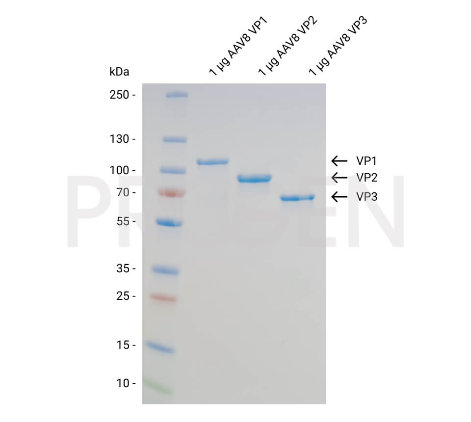
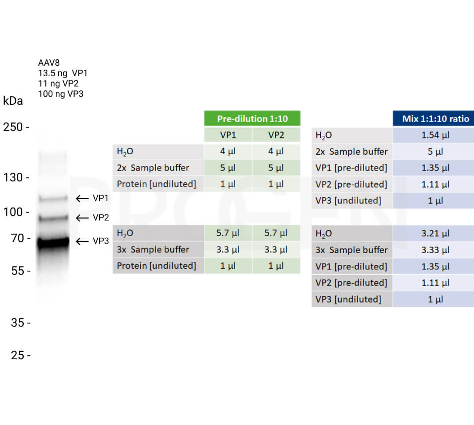
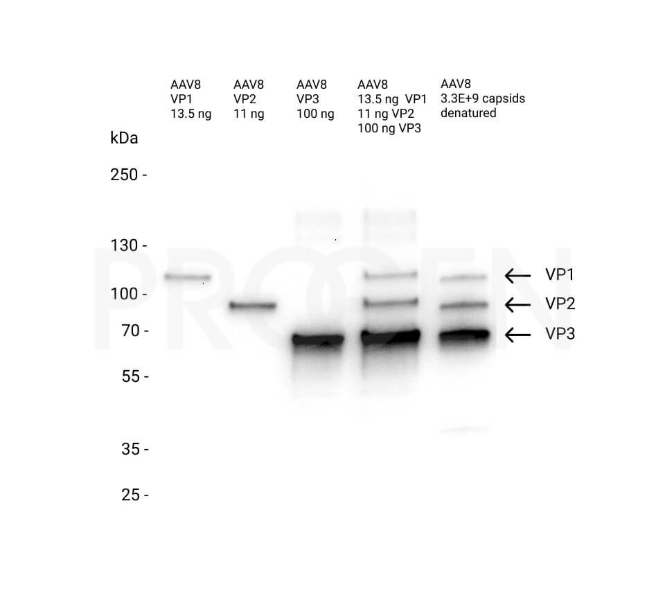
The recombinant AAV8 VP3 protein in combination with recombinant AAV8 VP1 (Cat. No. 640839) and recombinant AAV8 VP2 (Cat. No. 640840) can be used to create a mixture with the precise molar ratio of 1:1:10 to compare the protein composition of the viral capsid in your sample by protein detection methods, e.g. western blot. All three recombinant AAV8 capsid proteins are available as set (Cat. No. 72008) or as individual proteins (Cat. No. 640839, 640840, 640841).
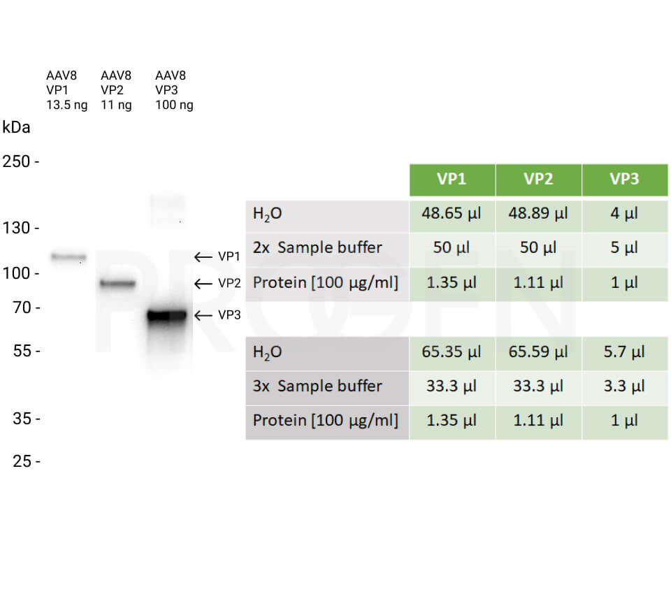
Note: please find an example how to prepare western blot samples in the pipetting scheme below. Aliquots of the remaining samples can be stored at -80°C for reuse.
Downloads
File
Category
Size
Filetype
Q & A's
There aren't any asked questions yet.
Customer Reviews
Login

