Epitope mapped
Highly published
Linear epitope
anti-AAV VP1/VP2/VP3 mouse monoclonal, B1, AFDye™ 488 Conjugate
Key Features
- AFDye™ 488 conjugate
- Mouse monoclonal
- Suitable for WB
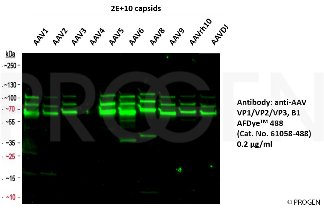
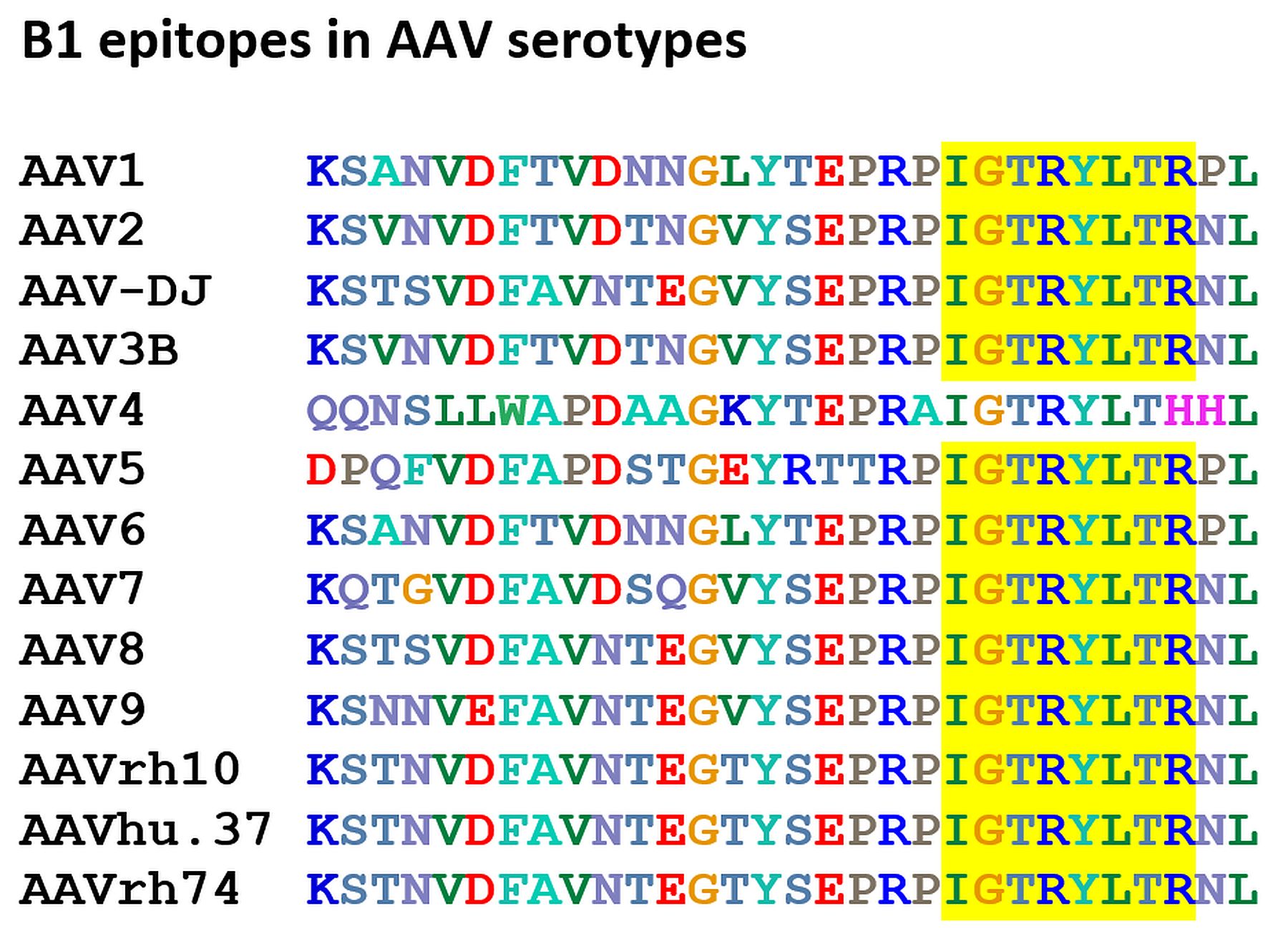
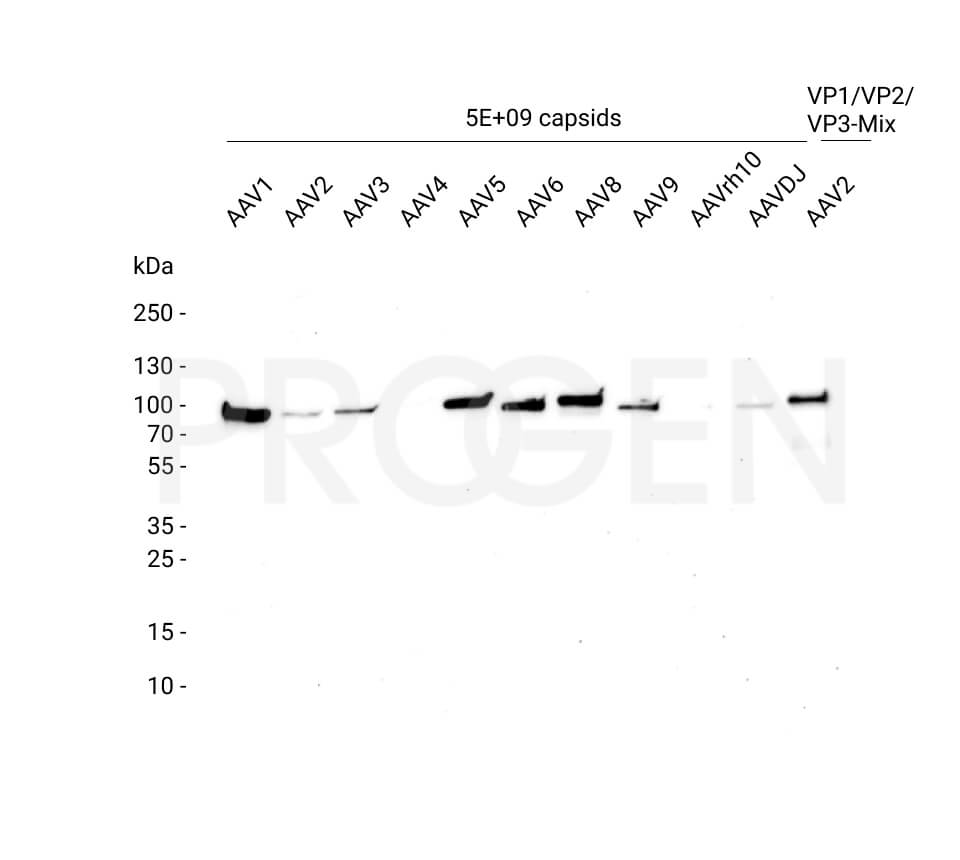
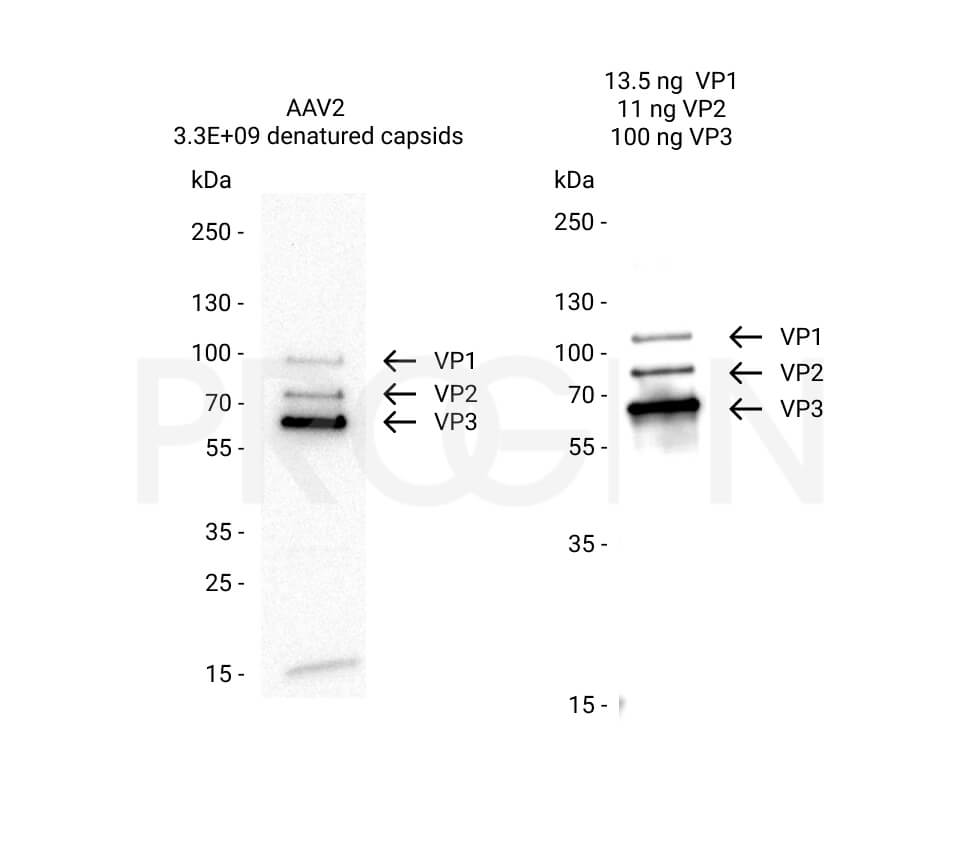
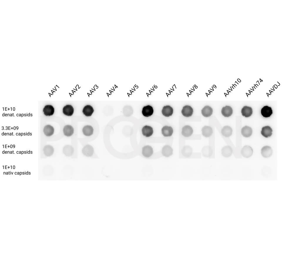
- Reacts with denatured VP1, VP2, VP3 proteins of AAV1, 2, 3, 5, 6, 7, 8, 9, rh10, DJ
- Isotype: IgG1
Product description
| Quantity | 50 µg (100 µl) |
|---|---|
| Antibody Type | Monoclonal |
| Host | Mouse |
| Isotype | IgG1 |
| Conjugate | AFDyeTM 488 |
| Application | WB |
| Purification | Affinity chromatography |
| Reactivity | AAV1, AAV2, AAV3, AAV5, AAV6, AAV7, AAV8, AAV9, AAVDJ, AAVrh10 |
| Storage | Up to 1 month: 2-8°C; long term storage in aliquots at -20°C; avoid freeze/thaw cycles |
| Intended use | Research use only |
| Clone | B1 |
| Immunogen | AAV2 capsids |
| Concentration | 500 µg/ml |
| Formulation | Liquid; PBS + 0.09% sodium azide |
Applications
| Tested applications | Tested dilutions |
|---|---|
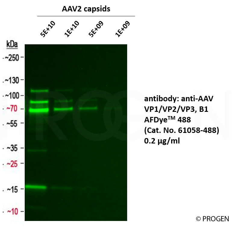
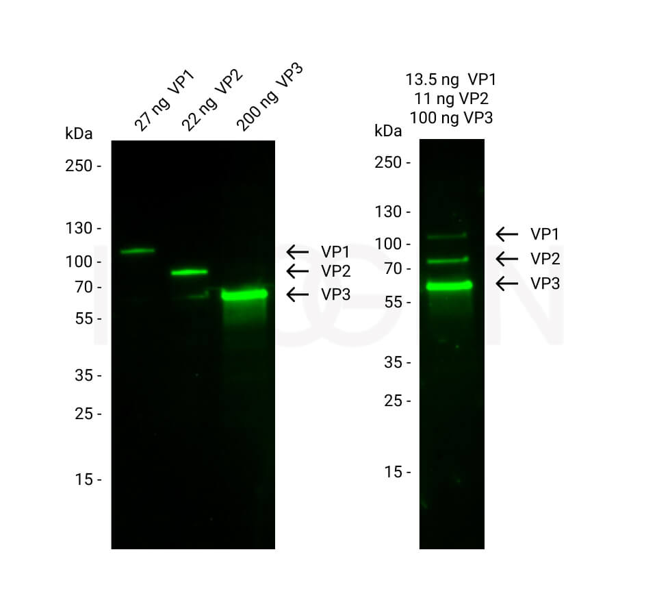
| Western Blot (WB) | 1:2,500-1:5,000 (0.1-0.2 µg/ml; detection limit 5E+09 capsids) |
Background
The B1 antibody reacts with free VP1, VP2 and VP3 of adeno-associated virus (AAV) and at a reduced degree with assembled viral particles. VP1 and VP2 are highly enriched in the nucleus, while non-assembled VP3 is evenly distributed in the nucleus and the cytoplasm. Epitope mapping experiments (Wobus et al., 2000) identified aa726 to aa733 (C-terminus; common to all 3 VP proteins) as the specific binding region. The antibody is also useful for characterization of different stages of infection.
The AFDyTM 488 is a green emitting dye, structurally and spectrally equivalent to Alexa FluorR 488 and therefore ideally suited for the 488 nm laser line (excitation/emission 494/517 nm). It can be used with a common FITC filter set.
The AFDyTM 488 is a green emitting dye, structurally and spectrally equivalent to Alexa FluorR 488 and therefore ideally suited for the 488 nm laser line (excitation/emission 494/517 nm). It can be used with a common FITC filter set.
Alexa FluorR is a registered trademark of Thermo Fisher Scientific.
Limited Use Label License: Research Use Only
Product is exclusively licensed to PROGEN Biotechnik GmbH. The use of these products for the development, manufacturing and sale of secondary products/derivatives which are based on the purchased products and/or which include the purchased product require a royalty based sub-license agreement.
References/Publications (42)
Publication
Species
Application
Species
AAV5, 9
Application
dot blot
Species
AAV9
Application
IHC/IF
Publication
Galibert, L. et al. Functional roles of the membrane-associated AAV protein MAAP. Sci. Rep. 11, (2021).
Species
AAV2
Application
WB
Species
AAV2
Application
WB
Species
AAV
Application
WB
Species
AAV1, 2, 3, 5, 6, 7, 8, 9, rh10
Application
dot blot
Species
human
Application
WB
Species
E. coli
Application
WB
Species
human
Application
WB
Species
human
Application
WB
Species
AAV2
Application
WB
Species
AAV2
Application
WB
Species
AAV8
Application
WB
Species
AAV2
Application
WB
Application
WB
Application
IP
Species
AAV9
Application
WB
Species
AAV5
Application
WB
Species
AAV8
Application
WB
Application
WB
Species
AAV2
Application
WB
Species
AAV1,AAV2,AAV3,AAV5,AAV6,AAV7,AAV8,AAV9
Application
dot blot
Species
AAV1,AAV2,AAV3,AAV5,AAV6,AAV7,AAV8,AAV9,AAVrh10
Application
WB
Species
AAV1,AAV2,AAV3,AAV5,AAV6,AAV7,AAV8,AAV9,AAVrh10
Application
WB
Species
AAV5
Application
WB
Species
AAV2
Application
WB,dot blot
Species
AAV1,AAV2,AAV5,AAV8,AAV9
Application
WB
Species
AAV5
Application
WB
Species
AAV2
Application
WB
Species
AAV2
Application
WB
Species
AAV1,AAV5,AAV6
Application
WB,ICC-IF
Species
AAV2
Application
WB
Species
AAV2
Application
WB
Species
AAV2,AAV5
Application
WB
Species
AAV2
Application
WB
Species
AAV2
Application
WB
Species
AAV2
Application
WB,ICC-IF
Species
AAV2
Application
epitope mapping
Species
AAV
Application
WB
Species
AAV2
Application
WB,IP,ICC-IF
Species
AAV2
Application
WB
Downloads
File
Category
Size
Filetype
Q & A's
There aren't any asked questions yet.
Customer Reviews
Login
FAQs
The concentration of unpurified supernatant, ascites, unpurified guinea pig serum and unpurified rabbit serum is not determined.
The concentration of purified antibodies is mentioned on the datasheet.
For prediluted antibodies the concentration may vary from lot to lot. The concentration of these antibodies is not mentioned on the datasheet and can be requested at support@progen.com.
The concentration of purified antibodies is mentioned on the datasheet.
For prediluted antibodies the concentration may vary from lot to lot. The concentration of these antibodies is not mentioned on the datasheet and can be requested at support@progen.com.
Most of our purified mouse antibodies contain 0.5% BSA as stabilizer. If BSA was added to the antibody solution, it is stated in the datasheet.
The supernatant format contains FCS proteins from cell culture medium supplemented with FCS.
The serum antibodies contain other proteins present in serum.
The supernatant format contains FCS proteins from cell culture medium supplemented with FCS.
The serum antibodies contain other proteins present in serum.
Please find the solvent for reconstitution and the amount of liquid for reconstitution on the antibody datasheet. The buffer formulation after reconstitution is also mentioned in the datasheet.
PROGEN offers different formats for mouse monoclonal antibodies. Different antibodies may not be offered in each format, the following are all possible formats:
- Supernatant and supernatant concentrate: This format contains hybridoma cell culture supernatant. The antibody is not purified and the antibody concentration is not determined. The antibody concentration may vary from lot to lot. Therefore we recommend to titrate the optimal concentration for the application used for each new lot.
- Lyophilized, purified: This format contains purified antibody in lyophilized form. The reconstitution of this antibody is described in the datasheet. The buffer composition after reconstitution is also mentioned on the datasheet.
- Liquid, purified: This format contains purified antibody in liquid format. The concentration is mentioned on the datasheet.
- Prediluted, purified: This format contains purified antibody in liquid format. Most antibodies in this format are diluted to be ready-to-use for IHC with standard tissue. But some antibodies of this format need further dilution for IHC. This is mentioned on the datasheet.
Lyophilized antibodies can be stored at 2-8°C until expiration.
Most of our liquid antibodies and reconstituted lyophilized antibodies may be stored for short term storage (up to 3 month) at 2-8°C. For long term storage we recommend to store the antibody at -20°C in aliquots. Please avoid freeze and thaw cycles.
Most of our conjugated antibodies should be stored at 2-8°C.
The individual storage conditions are mentioned on the datasheet.
The expiration date of our antibodies is indicated on the product label.
We offer bulk amounts starting from 1 mg for many of our mouse monoclonal or recombinant antibodies. Please contact us for further information or use the “bulk button” on the product page.
Most of our antibodies contain 0.09% sodium azide as preservative. If a preservative is added, it is mentioned in the datasheet.
The optimal antibody dilution for your specific protocol and application needs to be titrated in your lab with your equipment and sample. The optimal dilution may vary between protocols and samples. A good dilution for starting the titration is the dilution mentioned in the datasheet. If the sample needs a specific treatment (eg. Antigen retrieval for IHC on FFPE sections) this should also be mentioned on the antibody datasheet.
Anti-Adeno-Associated Virus Capsid Proteins Antibodies
For more information on the antibody binding epitopes of the capsid protein antibodies A1, B1 and A69 see the table below summarizing the published data from the following publication on the specific antibody binding sites:

Anti-Adeno-Associated Virus Particle Antibodies
For more information on the antibody binding epitopes on assembled AAV1, 2, 5, 8 and 9 capsids, see the tables below summarizing the published data from the following publications on the specific antibody binding sites: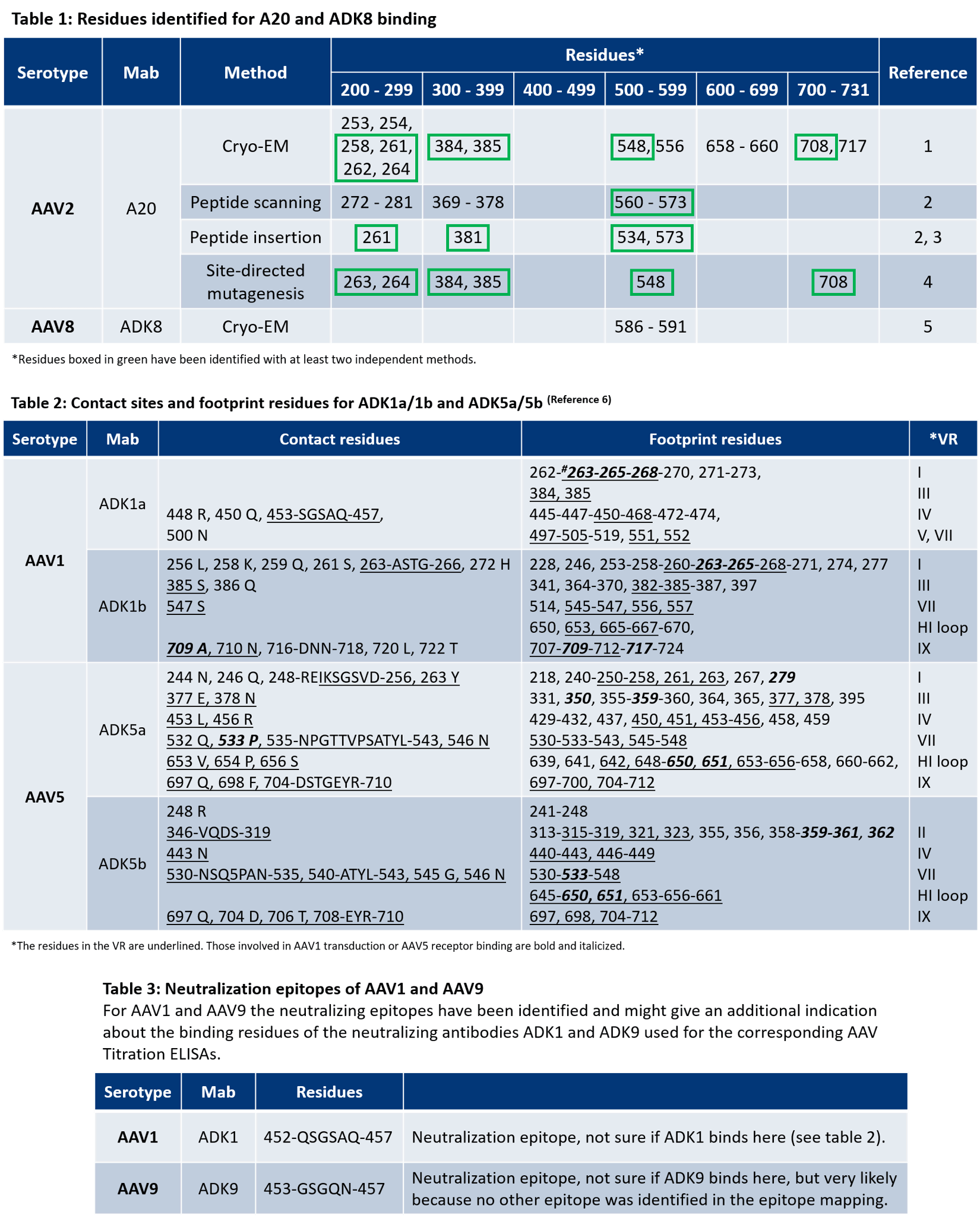
-
AAV 1
- Tseng, Y.-S. et al. Adeno-Associated Virus Serotype 1 (AAV1)-and AAV5-Antibody Complex Structures Reveal Evolutionary Commonalities in Parvovirus Antigenic Reactivity. J. Virol. 89, 1794–1808 (2015).
- McCraw et al. Structureof adeno-associated virus-2 in complex with neutralizing monoclonal antibody A20. Virology (2012) 431:40-9.
- Wobus, C. E. et al. Monoclonal antibodies against the adeno-associated virus type 2 (AAV-2) capsid: epitope mapping and identification of capsid domains involved in AAV-2-cell interaction and neutralization of AAV-2 infection. J. Virol. 74, 9281–93 (2000).
- Huttner et al. Genetic modifications of the adeno-associated virus type 2 capsid reduce the affinity and the neutralizing effects of human serum antibodies. Gene Ther (2003) 10:2139-47.
- Lochrie et al. Mutations on the external surfaces of adeno-associated virus type 2 capsids that affect transduction and neutralization. J Virol (2006) 80:821-34.
- Tseng, Y.-S. et al. Adeno-Associated Virus Serotype 1 (AAV1)-and AAV5-Antibody Complex Structures Reveal Evolutionary Commonalities in Parvovirus Antigenic Reactivity. J. Virol. 89, 1794–1808 (2015).
- Gurda, B. L. et al. Mapping a Neutralizing Epitope onto the Capsid of Adeno-Associated Virus Serotype 8. J. Virol. 86, 7739–7751 (2012).
EM structures of assembled AAV1, 2, 4, 5, 6, and 8 capsids with Fab-fragments are available in the following publications:
- Emmanuel, S. N. et al. Parvovirus Capsid-Antibody Complex Structures Reveal Conservation of Antigenic Epitopes Across the Family. Viral Immunolgoy. 34(1) (2021).
- McCraw, S. M. et al. Structure of adeno-associated virus-2 in complex with neutralizing monoclonal antibody A20. Virology. 432(1-2):40-9 (2012).
- Tseng, Y. et al. Adeno-Associated Virus Serotype 1 (AAV1)- and AAV5-Antibody Complex Structures Reveal Evolutionary Commonalities in Parvovirus Antigenic Reactivity. J Virol. 89(3):1794-1808 (2015).
- Jose, A. et al. High-Resolution Structural Characterization of a New Adeno-associated Virus Serotype 5 Antibody Epitope toward Engineering Antibody-Resistant Recombinant Gene Delivery Vectors. J Virol. 93(1):e01394-18 (2019).
- Silveria, M. A. et al. The Structure of an AAV5-AAVR Complex at 2.5 Å Resolution: Implications for Cellular Entry and Immune Neutralization of AAV Gene Therapy Vectors. Viruses. 12(11):1326 (2020).
PROGEN antibodies are shipped at ambient temperature. The antibodies are stable at ambient temperature for the shipment period. Please store the antibodies as indicated in the datasheet upon arrival.

