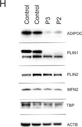
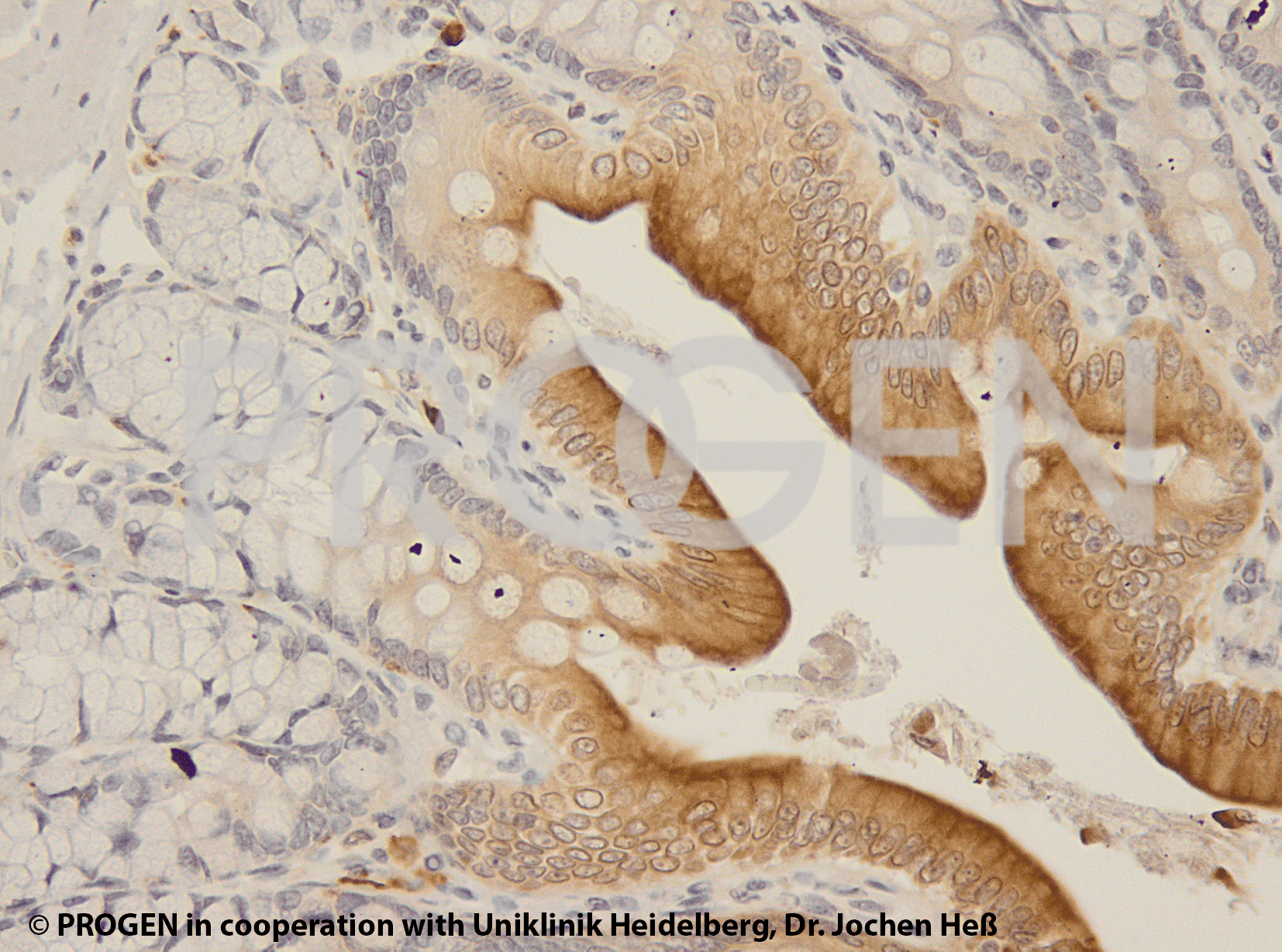
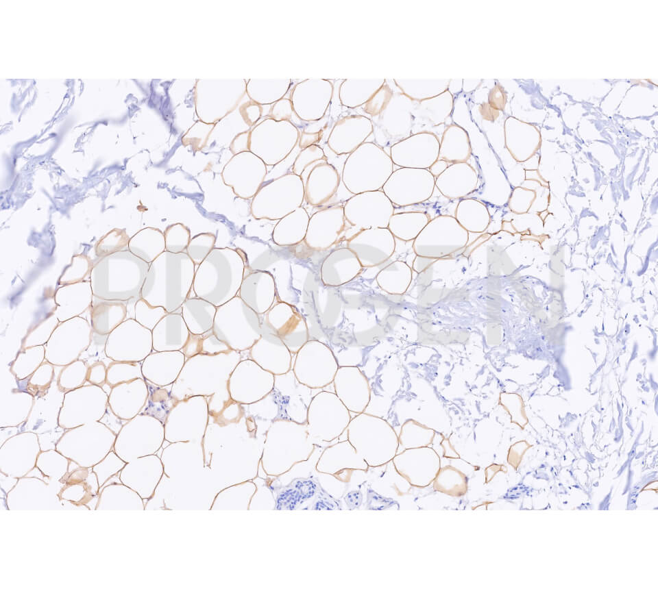
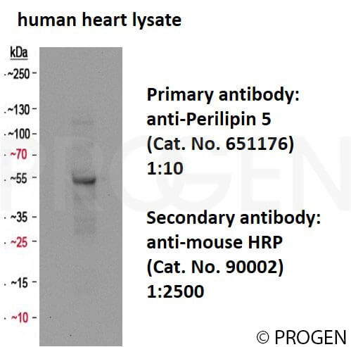
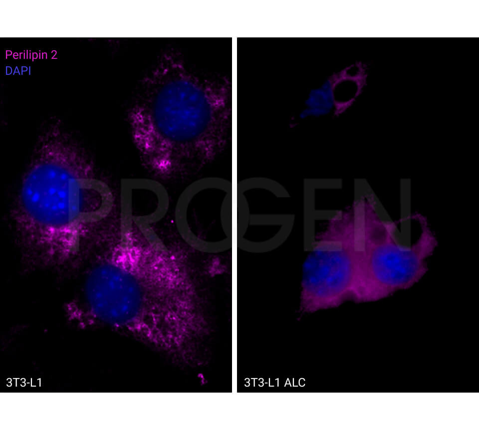
anti-Perilipin 2 (N-terminus aa 6-27) guinea pig polyclonal, serum
- Guinea pig polyclonal
- Suitable for ICC/IF, IHC and WB
- Reacts with human and mouse
Product description
| Quantity | 100 µl |
|---|---|
| Antibody Type | Polyclonal |
| Host | Guinea pig |
| Conjugate | Unconjugated |
| Application | ICC/IF, IHC, WB |
| Purification | Stabilized antiserum |
| Reactivity | Human, Mouse |
| Storage | Short term at 2-8°C; long term storage in aliquots at -20°C; avoid freeze/thaw cycles |
| Intended use | Research use only |
| Immunogen | Synthetic peptide (N-terminal aa 6-27 of human adipophilin / PLIN2) |
| Formulation | Contains 0.09% sodium azide and 0.5% BSA |
| UniprotID | Q99541 (Human), P43883 (Mouse) |
| Synonym | Perilipin-2, Adipophilin, Adipose differentiation-related protein, ADRP, PLIN2, ADFP |
| Note | Centrifuge prior to opening |
Applications
| Tested applications | Tested dilutions |
|---|---|
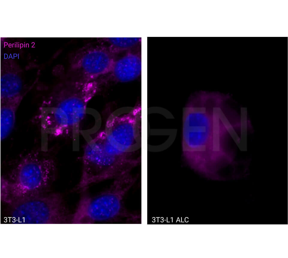
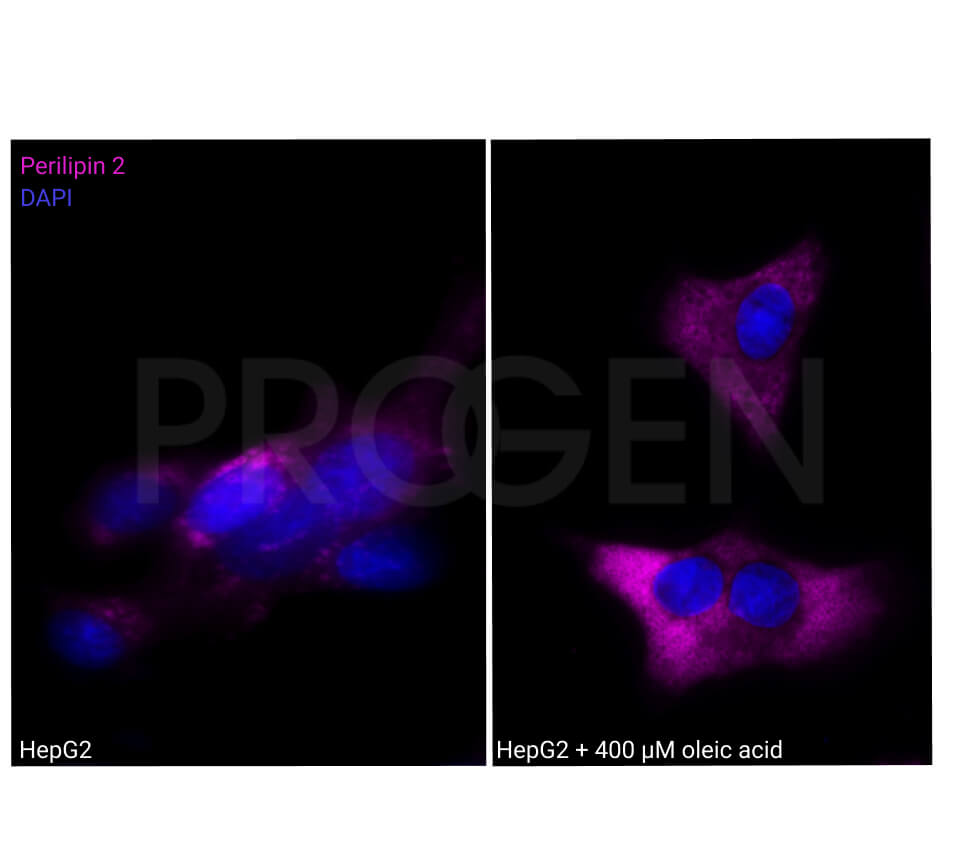
| Immunocytochemistry (ICC)/ Immunofluorescence (IF) | 1:50-1:100 |
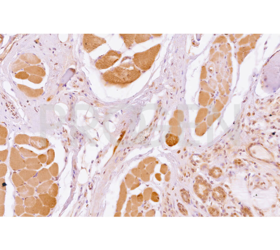
| Immunohistochemistry (IHC) - frozen | 1:100 |
| Immunohistochemistry (IHC) - paraffin | 1:100 (microwave treatment recommended) |
| Western Blot (WB) | 1:500-1:2,000 |
Background
Adipophilin / ADRP / PLIN2 is a ubiquitous component of lipid droplets. It has been found in milk fat globule membranes and on the surface of lipid droplets in various cultured cell lines (Heid et al. 1998; Targett-Adams et al. 2003); inducible by etomoxir.
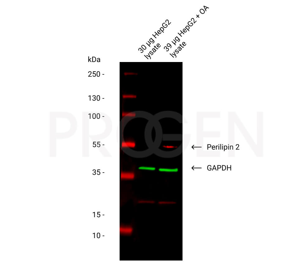
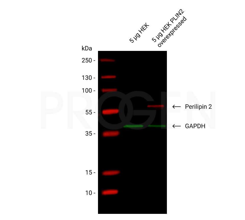
Enhanced expression of Adipophilin / ADRP / PLIN2 is a useful marker for pathologies characterized by increased lipid droplet accumulation. Such diseases include atheroma, steatosis, obesity and certain cases of liposarcoma. It also seems to be a potent marker for atherosclerosis. ADRP can also be used to study the virus entry of e.g. HCV via lipid droplets (Hope et al. 2002). Polypeptide reacting: Adipophilin / ADRP / PLIN2, MW 48,100 (calculated from aa sequence data); apparent Mr 52,000 (after SDS-PAGE); pI 6.72.
Tissue localization: Adipophilin / PLIN2 is positively detected in the glandular cells of lactating mammary gland (ductal cells are negative), zona fasciculata of the adrenal gland, Sertoli cells of the testis, and in fat-accumulating hepatocytes of alcoholic cirrhotic fatty liver; adipocytes are negative.
Also positively stained are lipid-storing CD 68-positive macrophages.
Reactivity on cultured cell lines: PLC.
Targett-Adams, P. et al. Live Cell Analysis and Targeting of the Lipid Droplet-binding Adipocyte Differentiation-related Protein. J. Biol. Chem. 278, 15998-16007 (2003).
Hope, R. G., Murphy, D. J. & McLauchlan, J. The Domains Required to Direct Core Proteins of Hepatitis C Virus and GB Virus-B to Lipid Droplets Share Common Features with Plant Oleosin Proteins. J. Biol. Chem. 277, 4261-4270 (2002).
Heid, H. W., Moll, R., Schwetlick, I., Rackwitz, H. R. & Keenan, T. W. Adipophilin is a specific marker of lipid accumulation in diverse cell types and diseases. Cell Tissue Res. 294, 309-21 (1998).
References/Publications (3)
Downloads
Q & A's
Customer Reviews
Login
FAQs
- PVDF membranes show better results than nitrocellulose (higher capacity, allows for more stringent washing conditions in case of background problems).
- Use freshly prepared blocking solution (e.g. 5% nonfat dry milk, 0.05% Tween 20), block for at least 1 h at room temperature.
- Use the antibody in a higher dilution, but prolong incubation time and exposure time.
- Always use a fresh aliquot of the antibody.
- Do not repeatedly freeze the antibody (eventually centrifuge shortly after thawing to remove cryo-precipitates).
- Include an additional washing step.
You might also try more stringent wash conditions, e.g. add 0.5 M NaCl to the wash buffer. - Always use a fresh aliquot of secondary antibody.
- In case you use ECL most the guinea pig antibody should be diluted further in order to get rid of the background.
The supernatant format contains FCS proteins from cell culture medium supplemented with FCS.
The serum antibodies contain other proteins present in serum.
- The fixation method used for lipidic material influences tremendously the quality of staining results.
Tissue fixation should not exceed 2% paraformaldehyde. - Heat induced antigen retrieval is recommended. In many cases a standard protocol using 10 mM citrate buffer (pH 6) works fine.
Most of our liquid antibodies and reconstituted lyophilized antibodies may be stored for short term storage (up to 3 month) at 2-8°C. For long term storage we recommend to store the antibody at -20°C in aliquots. Please avoid freeze and thaw cycles.
Most of our conjugated antibodies should be stored at 2-8°C.
The individual storage conditions are mentioned on the datasheet.
- Methanol/ acetone fixation: Immerse slide in precooled (-20°C) methanol for 5 min, immerse in precooled (-20°C) acetone for 30-60 sec, let specimen air dry before antibody incubation.
- Methanol/ acetone fixation plus detergent permeabilization: After methanol/ acetone fixation and air-drying dip slide either in a solution containing 0.1-0.2% Triton X-100 in PBS or in 0.1% saponin in PBS for 1-5 min at room temperature (enhances accessibility of many cytoskeletal antigens).
- Air-drying of the section.
- Block with the serum of the species in which the secondary antibody was raised for 30 min.
- Incubation with 1st antibody 1 h at RT in moist chamber.
- Wash 3x with PBS.
- Incubation with appropriate fluorescent secondary antibody, 30-60 min at RT.
- Wash 3x with PBS.
- Immerse shortly into ethanol.
- Let air dry.
- Cover with mounting medium.
- The use of milk preparations (skim milk, dry milk) for blocking and for antibody dilution buffer might be problematic. The antigen of the Perilipin 2 antibodies was originally found and described in bovine milk and some dry milk brands (or even different lots) might contain the epitopes and block it, thus abolishing the reaction with the antigen transferred onto the Western blot membrane. In this case Tween 20 or BSA or ovalbumin is recommended as e.g. blocking material.
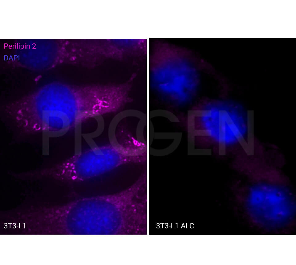
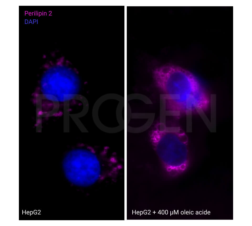
- Immune reaction with cultured cells: this depends on the age of the cell culture; old cells (more than confluent) tend to accumulate fat and are therefore positive for Perilipin 2. Freshly split cells contain only few lipid droplets and are not easily detectable as positive for Perilipin 2.
- Tissue with a high contents of fat might be problematic in electrophoretical separation. One has to load much material onto the SDS gel in order to get enough protein for a positive immunological signal. In addition the protein solubility is sometimes hindered by high fat concentration in the sample.
In guinea pigs the antibody concentration in serum varies from 10 to 20 mg/ml; specific antibodies represent normally about 0.1-1% of total IgG. Total protein concentration varies from 40 to 65 mg/ml, with the main constituent (about 60%) being albumin.

