Membranes

separation of the inside from the outside

The main functions of membranes are to form the shape and to regulate the exchange of substances from one side to the other through selective permeabilty. However, membranes also enable the messaging from one cell to another by the possibility to receive messages and process a proper response to a chmical or electrical signal.

In general, biological membranes consist of a lipid bilayer, a double sheet of different lipid molecules, called the phospholipid bilayer. In addition, specific membrane proteins and sugars are components of lipid bilayers. While membrane proteins help maintaining the structural integrity of membranes as well as its organization and the flow of materials, sugars are usually located on one of the sides of the bilayer and are bound to specific lipid or proteins. A biological membrane contains three types of lipids, phospholipids, glycolipids and sterols with each of the having a different properties. By adding sugars to lipids or proteins, these molecules can function as markers, e.g. some carbohydrate markers are present in diseases like cancer and can be used for diagnostics.  


Browse Membrane, Cytoskeleton & ECM Antibodies

Cytoskeleton

highly dynamic network maintaining  fundamental requirements

The main function of the cytoskeleton is to from and maintain the shape of the cell and support the resitance to deformation. It further is connected with the extracellular cennective tissue and therefore has a stabilizing function for entire tissues. By controlled deformation due to contraction, the cytoskeleton also allows the migration of cells. However, also involvment in signaling pathways within the cells and uptake of specific components, e.g. via endocytosis as well as the division of chromosomes during cell cycle are included in the overall function of the cytoskeleton.

The complex and highly dynamic network of protein filaments streches from the cell nuclear to the cellular membrane and is mainly composed of three structures, the microfilaments, intermediate filaments and microtubules. All structures enable fast disassembly and growth to maintain the essential functions of the cytoskeleton.

Extracellular Matrix (ECM)


Extracellular molecules such as proteins and glycosaminoglycans (GAGs) and minerals form a three-dimensional network providing structural support to surrounding cells. Collagen, enzymes, glycoproteins and hydroxyapatite are common molecules of the ECM. The main function is cell adhesion, provision of cell-to-cell communication and differentiation. However, its composition varies strongly between different multicellular structures.

The components of the ECM are produced and secreted by the surrounding cells and integrate to the existing matrix. Fibronectin and Lamin are examples for cell adhesion proteins within the ECM, however the ECM can also connect with actin filaments or hemidesmosomes of the cells.

Browse Membrane, Cytoskeleton & ECM Antibodies

Desmosomes

and their role in cell adhesion

To maintain physical and mechanical integrity of cells that are extremely exposed to mechanical stress, desmosomes have an essential role. Therefore, desmosomes are prominently distributed in epithelial and cardiac muscle cells. Desmosomes belong to the epithelial junctional complex which further include tight junctions and adherens junctions. Moreover, it is described as a cadherin-mediated junctional procedure that mechanically couples the intermediate filaments of neighbored cells. Further, the transmembrane region and the plaque region that anchors the intermediate filaments are components of the two symmetrical halves that are forming the desmosome-intermediate filament complex (DIFC).

Loss of function mutations in the desmosomal protein complex have severe consequences to human pathologies including skin blistering upon mechanical stress, lethality prior to gastrulation or heart muscle dysfunction.

The desmosome-intermediate filament complex (DIFC)

  • network of non-classical cadherins, linker proteins and intermediate filaments
  • divided into three main regions
  • the extracellular core region, containing desmoglein and desmocollin
  • the outer dense plague containing intracellular ends of desmoglein and desmocollin as well as N-terminal sides of desmoplakin, plakoglobin and plakophilin
  • the inner dense plaque containing C-terminal ends of desmoplakin and their attachement to intermediate filiaments


Browse Desmosome Antibodies

Protein family and Research Antibodies

PROGEN offers a comprehensive portfolio, containing different antibodies against all desmosome proteins. Our desmosome antibodies are highly published and have been independently validated for relevant applications (western blot, IHC, IF and ICC). Protocols and data provide useful information for your experimental design.


The family of desmosomal cadherins comprise three variants of desmocollin (DSC1-3) and four variants of desmoglein (DSG1-4). Both are coupling adjacent cells together by the formation of homo- and heterophilic trans-interactions. Furthermore, the desmosomal cadherins interact intracellularly with the linker armadillo proteins that are associated to desmoplakin. Additionally, the transcellular adherence between desmocollin and desmoglein is Ca2+ dependent.
Generally, desmosomal cadherins are regulating normal physiological processes such as differentiation and epithelial morphogenesis.


Browse Desmocollin Antibodies Browse Desmoglein Antibodies

The armadillo proteins comprise plakoglobin (PG/JUP) and three variants of plakophilin (PKP1-3) that are located at the cell junctions. There are multiple and differentiation dependent isoforms of the armadillo proteins and of the desmosomal cadherins, which is associated with differences in desmosome composition. The expression of plakophilin is cell-type specific and the protein is located at the cell border supporting desmosome assembly and stability. Moreover, plakophilins interact with all major desmosomal structures and cytoskeletal components like keratin. Therefore, they are described as desmosomal crosslinkers. Loss of function mutations in PKP cause a reduction in size and number of the desmosomes and lead to an increase in migration. Because of that, mutations of PKP are implicated in human genetic disorders and cancer.
Plakoglobin promotes cell adhesion by interacting with desmosomal cadherins. More precisely, plakoglobin binds to the intracellular catenin-binding region of desmosomal cadherins and forms a bridge between the cadherin tail and the intermediate filament binding proteins like desmoplakin.
Furthermore, plakoglobin is an important component contributing to normal skin physiology and has important roles in tissue integrity and cell signaling.

Browse Plakoglobin Antibodies Browse Plakophilin Antibodies

Desmoplakin (DSP) has two major isoforms DPI and DPII and is a member of the plakin family of cytolinker proteins. Moreover, it is an essential component of the desmosome due to its role in linking the desmosomal plaque to the intermediate filament. The loss of DP leads to a drastic reduction of desmosomes and thus has critical roles in skin and heart tissues.

Browse Desmoplakin Antibodies


IHC Images

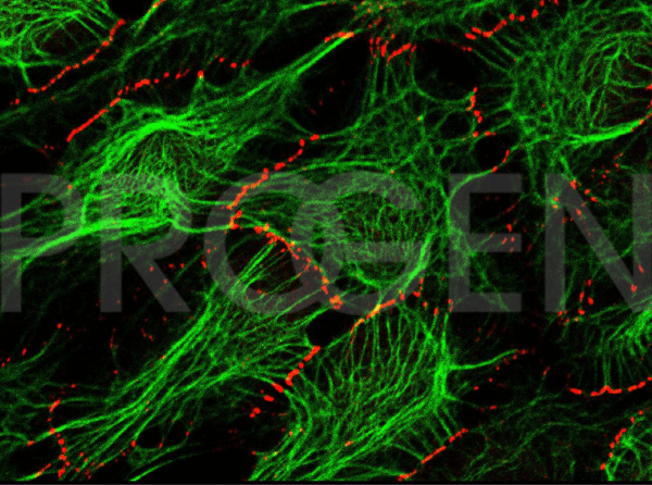
HaCaT Plakophilin-1 (red), cytokeratin (green) (courtesy of L. Langbein)

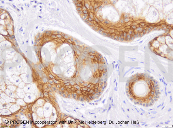
IHC of human skin using anti-Desmoglein 1/2 (Cat. No. 61002) (courtesy of J.Heß, University Hospital Heidelberg)

Publications

  1. Nekrasova, O. et al. Desmosomal cadherin association with Tctex-1 and cortactin-Arp2/3 drives perijunctional actin polymerization to promote keratinocyte delamination. Nat.Commun. 9, 1053 (2018).
  2. Vielmuth, F. et al. Keratins Regulate p38MAPK-Dependent Desmoglein Binding Properties in Pemphigus. Front.Immunol. 9, 528 (2018).
  3. Price, A. et al. Mechanical loading of desmosomes depends on the magnitude and orientation of external stress. Nat.Commun. 9, 5284 (2018).
  4. Shafraz, O. et al. E-cadherin binds to desmoglein to facilitate desmosome assembly. Elife. 7, (2018).
  5. Kudo, I. et al. Particular gene upregulation and p53 heterogeneous expression in TP53-mutated maxillary carcinoma. Oncol.Lett. 14, 4633-4640 (2017).
  6. Izawa, G., Kobayashi, W., Haraguchi, M., Sudo, A. & Ozawa, M. The ectopic expression of Snail in MDBK cells does not induce epithelial-mesenchymal transition. Int. J. Mol. Med.36, 166–72 (2015).
  7. Homberg, M. et al. Distinct Impact of Two Keratin Mutations Causing Epidermolysis Bullosa Simplex on Keratinocyte Adhesion and Stiffness. J. Invest. Dermatol. 135, 2437–2445 (2015).
  8. Yang, C. et al. Plakophilin 1-deficient cells upregulate SPOCK1: implications for prostate cancer progression. Tumour Biol. 36, 9567–77 (2015).
  9. Fischer-Kešo, R. et al. Plakophilins 1 and 3 bind to FXR1 and thereby influence the mRNA stability of desmosomal proteins. Mol. Cell. Biol. 34, 4244–56 (2014).
  10. Dayal, J. H. S. et al. Type VII collagen regulates expression of OATP1B3, promotes front-to-rear polarity and increases structural organisation in 3D spheroid cultures of RDEB tumour keratinocytes. J. Cell Sci.127, 740–51 (2014).
  11. Rickelt, S. Plakophilin-2: a cell-cell adhesion plaque molecule of selective and fundamental importance in cardiac functions and tumor cell growth. Cell Tissue Res. 348, 281–94 (2012).
  12. Jennemann, R. et al. Loss of ceramide synthase 3 causes lethal skin barrier disruption. Hum. Mol. Genet. 21, 586–608 (2012).
  13. Resnik, N. et al. Desmosome Assembly and Cell-Cell Adhesion Are Membrane Raft-dependet Processes. Cell Biology 286, 1499-1507 (2011).
  14. Broussard, J. et al. Desmosome regulation and signaling in disease. Cell Tissue Res. PupMed. 360,501-12 (2019).
  15. Kowalcyk, A. et al. Structure, function, and regulation of desmosomes. Prog Mol Bio Transl Sci. PupMed. 116,95-118 (2013).

Top Selling Antibodies

anti-Lamin B mouse monoclonal, X223, lyophilized, purified
Cat. No: 61047C
  • Mouse monoclonal
  • Suitable for ICC/IF, IHC and WB
  • Reacts with bovine, human, mouse, rat, rat kangaroo and trout
  • Isotype: IgG1

$251.00*
anti-Desmoglein 2 mouse monoclonal, 10G11, lyophilized, purified
Cat. No: 61059_1
  • Mouse monclonal
  • Suitable for IHC and WB
  • Reacts with human
  • Isotype: IgG1

Variants from $120.00*
$304.00*
anti-Desmoglein 1/2 mouse monoclonal, DG 3.10, lyophilized, purified
Cat. No: 61002
Quantity: 50 µg
  • Purified, lyophilized
  • Mouse monoclonal
  • Suitable for ICC/IF, IHC and WB
  • Reacts with bovine, human, rat
  • Isotype: IgG1

Variants from $120.00*
$304.00*
anti-Desmoplakin 1 mouse monoclonal, DP-2.17, lyophilized, purified
Cat. No: 61024
  • Mouse monoclonal
  • Suitable for IHC and WB
  • Reacts with bovine, chicken, human, mouse and rat
  • Isotype: IgG1

$304.00*
anti-Desmocollin 3 mouse monoclonal, Dsc3-U114, lyophilized, purified
Cat. No: 61093
Quantity: 50 µg
  • Purified, lyophilized
  • Mouse monoclonal
  • Suitable for IHC and WB
  • Reacts with human, mouse, rat
  • Isotype: IgG1

Variants from $96.00*
$251.00*

Know what you need?

Login and order directly
from your account.

Order Now

Need more information?

Visit our support hub for FAQs,
references and downloads.

Visit Support Hub