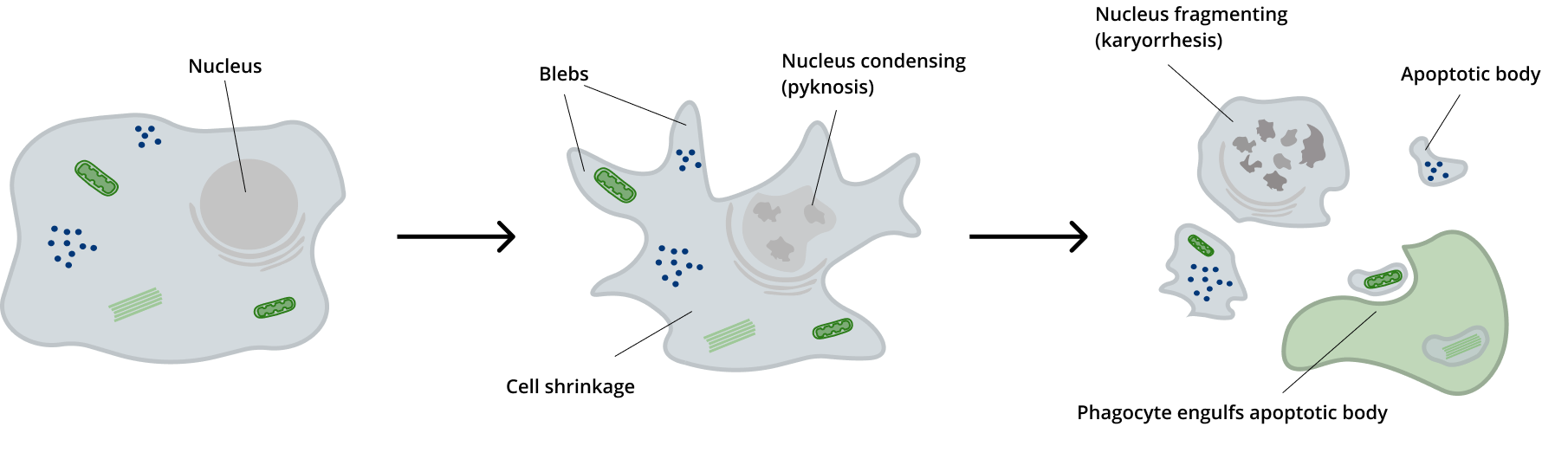
Autophagy & Apoptosis
Know what you need?
Order Now


 guinea pig polyclonal, serum")
anti-p62/ SQSTM1 (C-terminus) guinea pig polyclonal, serum
$343.00*
Cat. No: GP62-C
- Guinea pig polyclonal
- Suitable for IHC and WB
- Reacts with bovine, human, mouse and rat

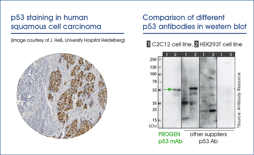
anti-p53 Protein mouse monoclonal, Bp53.11, lyophilized, purified
$265.00*
Cat. No: 61039
Quantity:
50 µg (2x25 µg vials)
- Purified, lyophilized
- Mouse monoclonal
- Suitable for ICC/IF, IHC and WB
- Reacts with human
- Isotype: IgG2a
Variants from $96.00*